18F-FC505
Vulnerable Atherosclerotic Plaque
Vulnerable atherosclerotic plaque (atherosclerotic plaque) is a vascular disease with formation of atheroma due to deposition of cholesterol on the lining of blood vessels and endothelial cell proliferation. The inside of the atheroma becomes like porridge and its neighboring area is surrounded by thin fibrous membrane. When the plaque becomes unstable, it ruptures, causing thrombus to form in the blood vessels. If blood vessels are blocked by thrombus, this causes serious diseases depending on the site of blockage. If cerebral blood vessels are blocked, stroke (cerebral infarction, cerebral hemorrhage) may occur; if coronary artery is blocked, myocardial infarction may occur; and if limb vessels are blocked, peripheral vascular disease may occur. These atherosclerotic plaques do not show any symptoms when the degree of narrowing is not severe, but sudden death or severe disability could occur when ruptured.

            Cerebrovascular diseases and cardiovascular diseases are caused primarily by atherosclerosis and are the leading cause of mortality worldwide. In Korea Cerebrovascular diseases and cardiovascular diseases rank the 2nd and 3rd cause of death after cancer, respectively. Currently, the diagnostic methods for these vascular diseases are carotid ultrasonography, abdominal ultrasonography, CT, coronary calcification test, and others. However, these diagnostic methods only confirm the degree of anatomical stenosis of blood vessels and cannot distinguish the histological characteristics of plaques. Stable plaques are less likely to rupture and are less likely to be related to cardiovascular diseases. Biopsy is required to confirm whether the plaque formed on the blood vessel is stable or not (Figure 1). Because acute diseases such as stroke and myocardial infarction should be judged by the histological characteristics of the plaque rather than by the stenosis of the blood vessels, PET radiopharmaceutical that can identify the characteristics of the plaque.

Histological Features of Atherosclerotic Plaques and Apoptosis
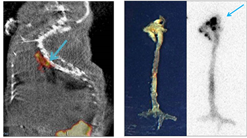
Various biological events occur in atherosclerotic plaques. Representative events include inflammation, angiogenesis, apoptosis, hypoxia, calcification and others that include lipid rich core. In particular, apoptosis is an active event in atherosclerotic plaques. Thus, the apoptosis-targeting substances may have specificity in atherosclerotic plaques.


		ApoPepApoPep is a peptide consisting of six amino acids that bind to apoptotic cells, and was developed by a phage display method in Kyungpook National University School of Medicine. The protein that ApoPep binds was found to be Histone 1 (H1), which is located in the cell nucleus in normal cells, but migrates to the cell membrane surface upon apoptosis. Since ApoPep contains a cysteine residue and has a disadvantage of being easily modified, structurally more stable cyclic ApoPep (cApopep) has been synthesized, and confirmed to have increased stability. In addition, it was confirmed that the cyclic Apopep compound had slightly increased binding ability to apoptotic cell

		18F-ApoPep-118F-ApoPep labeled with 18F at the amine end of ApoPep was administered to atherosclerotic plaque model mice (ApoE -/-), and MicroPET/CT image was acquired. As a result, it was confirmed that the uptake was relatively small but selectively bound to the aortic arch and the descending aorta in which many atherosclerotic plaques were generated. In terms of pharmacokinetics, 18F-ApoPep was rapidly excreted through kidneys.


		18F-FC505An improved version of 18F-ApoPep was designed and synthesized which is named as 18F-FC505. 18F labeling yield and pharmacokinetic properties were improved based on stable cyclic ApoPep. Atherosclerotic plaque model mice (ApoE -/-) were administered with 18F-FC505 to obtain MicroPET/CT image. It showed that 18F-FC505 selectively binds to atherosclerotic plaques formed in the aortic arch and the descending aorta. In addition, it was rapidly excreted through renal system.